Baby at 9 weeks’ gestation
Week 9 of pregnancy is the second week of the third trimester. It is still in the early stages of pregnancy.
The weeks and months of pregnancy are counted in “full” numbers from week 0, whereas the months of pregnancy are counted in “count” numbers from month 1. The third trimester is therefore weeks 8-11 of pregnancy.
Baby condition
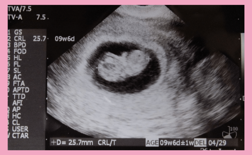
At 9 weeks’ gestation, the baby’s size (foetal cephalic length: CRL) is 20-30 mm, the gestational sac (GS) is 5.7 cm and the weight is around 1-3 g. It is just the size of a strawberry. The foetus is developing into a human-like shape, with a separation and a head and body. This is also the time when the face, fingers and toes and other small parts of the body are gradually being formed. The reproductive organs will also begin to develop around 9 weeks’ gestation.
The foetal heartbeat is also at its fastest at 9 weeks’ gestation. The foetal heart rate is around 90-100 beats per minute at 5 weeks’ gestation, which is often the first time it is confirmed by ultrasound at this time. The heart rate increases steadily until the ninth week of pregnancy, peaking at 170-180 beats/minute in the middle of the ninth week. Thereafter, the foetal heart rate gradually decreases as the gestational weeks progress, stabilising at around 150 beats/min around 16 weeks’ gestation until the day of delivery.

Echocardiogram at 9 weeks’ gestation
When the baby’s gender is known
Mothers may be concerned about the gender as well as the health of their baby. Many mothers cannot wait to find out when they will know the sex of their baby.
Echocardiography cannot determine the sex of a small baby at nine weeks’ gestation. The sex of the baby is determined by the sex chromosomes. If it is a boy, it will have an XY chromosome; if it is a girl, it will have an XX chromosome.
NIPT at Hiro Clinic NIPT can determine the sex of the baby by testing the sex chromosomes. NIPT can be performed from the early stages of pregnancy, allowing the sex of the baby to be known at an early stage.
Maternal changes that appear at 9 weeks’ gestation
In the mother’s body at 9 weeks’ gestation, the amount of hormones produced during pregnancy (hCG) increases and the body changes that are typical of pregnant women gradually arrive.
Maternal changes and morning sickness
At around 9 weeks’ gestation, the waist begins to enlarge, as is typical of pregnant women. The breasts also begin to enlarge due to hormonal effects.
This is also when morning sickness peaks. Many mothers report feeling sick all day long and worrying about malnutrition due to morning sickness. There is no medicine to control morning sickness, but it is believed that morning sickness symptoms become less severe around 12 weeks’ gestation and subside around 16 weeks’ gestation. There is no need to force yourself to eat when morning sickness is difficult. It is important to take care not to become dehydrated and to stay at rest.
Problems likely to occur and what to look out for
Weeks 7-9 of pregnancy are said to be the period when the probability of miscarriage is highest. However, most miscarriages that occur during this period are caused by chromosomal abnormalities and are unlikely to be caused by problems in the pregnant woman’s life or behaviour.
As the placenta has not yet stabilised, it is not uncommon for an imminent miscarriage to occur as a result of excessive work or exercise.
This is also the time when morning sickness symptoms reach their peak. Morning sickness symptoms vary from person to person, with some having mild morning sickness and others having morning sickness so severe that it is difficult to drink fluids. There are no medications to control morning sickness, so it is important to find ways to relieve it. However, severe dehydration can be dangerous for both mother and child, and early medical attention should be sought if there is a lack of fluids or significant weight loss.

Examination at 9 weeks’ gestation
The main antenatal check-ups carried out at 9 weeks’ gestation are listed below. In addition to publicly funded tests, there are also various self-funded tests that you may wish to consider if necessary. It is difficult to detect chromosomal abnormalities such as 21 trisomy (Down syndrome) in babies at an early stage during general antenatal check-ups.
Prenatal checkup
Pregnancy check-ups are important regular check-ups to protect the health of mother and child during pregnancy and to examine the baby’s growth. According to the Ministry of Health, Labour and Welfare, it is recommended to have a check-up once every four weeks from early pregnancy to 23 weeks, once every two weeks from 24 weeks to 35 weeks and once a week from 36 weeks to delivery. Pregnancy check-ups are not covered by insurance, but you can apply for subsidies from your local government, so make sure you check with your local office when you receive your Maternal and Child Health Handbook.
1.Assessing health status
Medical interview and examination
2.Inspection and measurement
his is to check the health of the pregnant woman and the development of the baby. Blood pressure, oedema (swelling), urine test (sugar and protein), weight and height (first check-up only). Maternal weight is considered a particularly important measurement point, and women who are underweight (BMI <18.5) are at increased risk of imminent or premature birth.
Women who tend to be obese (BMI≥25), on the other hand, are at increased risk of gestational hypertension, gestational diabetes, caesarean section deliveries and huge babies. The risk of other birth defects (congenital diseases), such as stillbirth and spina bifida, is also increased.
3.Health guidance
The service includes advice on diet and health, as well as consultation on concerns and worries about pregnancy, childbirth and childcare.
4.Medical tests performed as necessary
4-1.Blood test
- Items performed once initially: blood group (ABO blood group, Rh blood group, irregular antibodies), blood count, blood sugar, hepatitis B antigen, hepatitis C antibody, HIV antibody, syphilis serological reaction, rubella virus antibody.
- One test before 30 weeks’ gestation: HTLV-1 antibodies
The above tests are designed to ensure a safe childbirth: hepatitis B, hepatitis C, HIV, syphilis and HTLV-1 can be transmitted from mother to child, so if a positive test is confirmed, discuss treatment with your doctor.
4-2.Cervical cancer screening (cytology)
Once in early pregnancy
4-3.Ultrasound scan
Twice before 23 weeks’ gestation. If the date of ovulation cannot be determined, the cephalic length (CRL) at 8-10 weeks’ gestation is measured by echocardiography and used to estimate the expected date of delivery. The test is also considered a prenatal diagnosis in the broadest sense, as it provides a direct view of the baby in the abdomen.
4-4.Genital chlamydia testing
The test is performed once before 30 weeks’ gestation to prevent mother-to-child transmission. If the test is positive, the baby is treated with antibiotics to prevent infection.
4-5.GBS test
Test for GBS (group B haemolytic streptococci) at 35-37 weeks’ gestation. If positive, antimicrobials are administered before delivery as it is a rare cause of neonatal pneumonia and septicaemia.
Self-funded tests
1.Bacterial culture test
This is a test to check whether the vaginal flora is normal and free from bad bacteria. The presence of bad bacteria can cause an imminent miscarriage or premature birth.
2.TPHA test
Syphilis testing. The syphilis serological reactions available at antenatal check-ups rarely result in false positives (a positive result when the test is actually negative) in pregnant women; combining the TPHA test is considered to be a more accurate way of determining this.
3.Toxoplasma antibody test
This test is required for pet owners, especially cats. Infection during pregnancy can often lead to congenital toxoplasmosis in the baby, causing chorioretinitis, calcification of the brain and hydrocephalus. If infection is detected, treatment of the mother during pregnancy can prevent transmission to the baby.
4.Breast cancer checkup
Breast cancer progresses quickly during pregnancy, so if you have not had a breast cancer check-up within the past year, it is advisable to have a check-up early in your pregnancy.
5.NIPT
NIPT can be carried out from early pregnancy using only maternal blood. By collecting and analysing the baby’s DNA from the mother’s blood, the test is designed to determine the risk of chromosomal abnormalities in the foetus, such as 21 trisomy (Down syndrome)・18 trisomy (Edwards syndrome)・13 trisomy (Patau syndrome).
At Hiro Clinic NIPT, you can make an appointment for NIPT when your pregnancy is confirmed by ultrasound examination.
When can Down syndrome be detected by ultrasound?
A mother’s biggest concern during pregnancy is the health of her baby. At around 9 weeks’ gestation, the baby’s heartbeat can be clearly seen by ultrasound. However, the baby is still very small at this time and an echo scan cannot detect conditions such as 21 trisomy (Down syndrome).
An ultrasound scan generally reveals a 21 trisomy (Down syndrome) after 11 weeks of pregnancy. During the echo examination, the baby’s occipital and neck swelling is visually checked. The swelling in the back of the head and neck is expressed as a numerical value called the ‘NT value’ (=Nuchal Translucency), and since the NT value tends to gradually thicken even in normal babies, it is impossible to detect 21 trisomy (Down syndrome) or congenital disorders caused by other chromosomal abnormalities by echo examination alone. Therefore, it is not uncommon for a baby’s congenital condition that was not detected during pregnancy to be discovered after birth.
Chromosomal abnormalities in babies detected by NIPT
Congenital disorders due to chromosomal abnormalities in the baby that cannot be detected during antenatal check-ups by echocardiography. Detecting chromosomal abnormalities in babies at an early stage of pregnancy is very important for a healthy pregnancy, as it helps to identify the risk of miscarriage.
NIPT is a test that can be performed early in pregnancy. A blood sample is taken from the mother’s arm to check the baby’s risk of congenital heart disease due to chromosomal abnormalities.
NIPT does not cause direct damage to the baby and can detect 21 trisomy (Down syndrome) with a high accuracy of 99.9% in both sensitivity and specificity.
For mothers who have difficulty going out due to the peak of morning sickness, Hiro Clinic NIPT offers an internet-based system for booking appointments, consent forms and medical questionnaires. Please contact Hiro Clinic NIPT for advice.
【References】
- Ministry of Health, Labour and Welfare – Maternal Health Examination
- SRL comprehensive inspection guide – Syphilis Quantitative TPHA
Article Editorial Supervisor

Dr Hiroshi Oka
NIPT specialist clinic, MD
Graduated from Keio University, School of Medicine
 中文
中文


