- Is it true that Down syndrome can be detected by ultrasound (echo photo) at 7 weeks pregnant?
- About Down syndrome
- Characteristics of Down syndrome visible on ultrasound (echo photography)
- If Down syndrome is suspected based on ultrasound images, NIPT (New Prenatal Diagnosis) is performed
- Probability of developing Down syndrome by age group
- If your baby is diagnosed with Down syndrome
- まとめ
Is it true that Down Syndrome can be detected with an ultrasound (ultrasound photo) during the 7th week of pregnancy?
Unfortunately, it is not true that Down Syndrome can be detected by ultrasound at seven weeks of pregnancy .
In the first place, ultrasound (ultrasound photography) can only observe the characteristics of Down syndrome , but it cannot definitively diagnose that the baby has Down syndrome . It is said that the earliest that ultrasound (ultrasound photography) can diagnose the characteristics of Down syndrome is after the 11th week of pregnancy.
About Down Syndrome
First of all, what kind of disease is Down Syndrome ? Let’s review the correct information about Down Syndrome .
What is Down Syndrome?
The official name for Down syndrome is ” Down syndrome .”
Down syndrome (trisomy 21) is a chromosomal abnormality that occurs when there is an extra 21st chromosome, and is the most common genetic disorder in newborns. Down syndrome is named after Dr. John Langdon Down, who first reported it.
Because this is an abnormality in chromosomes within cells, it often affects the development of muscles and internal organs, so support must be provided at an early stage to correct developmental delays.
Characteristics of Down Syndrome
In addition to a distinctive appearance, Down syndrome can also be accompanied by delayed intellectual and physical development and other illnesses.
Down Syndrome Physical Characteristics
Babies with Down’s syndrome have many distinctive features, including a small head with a flat back, a wide, flat face, especially slanted eyes and a low-set nose, excess skin at the back of the neck, and small, round, low-set ears.
The fingers are short, the pinky has only two joints instead of the usual three and is bent inward, there is often only one horizontal crease on the palm, and fingerprints often have a horizontal line rather than a circular pattern.
Another characteristic is that they are generally short in stature and at higher risk of becoming obese.
Down Syndrome Intelligence and Mental Characteristics
Intelligence quotients (IQs) vary from person to person, so it is difficult to generalize, but if the IQ of a normal child is 100, the average IQ of a child with Down syndrome is about 50.
In terms of behavior, attention deficit, hyperactivity, and autistic behavior (such as repeating the same thing over and over again) are often observed, and it is known that there is a relatively high risk of depression.
Down Syndrome Other characteristics
Approximately half of people with Down syndrome have heart disease. The most common is a condition in which the walls separating the heart chambers do not form properly (endocardial cushion defect, ventricular septal defect, etc.).
In addition, there are many cases of digestive system diseases such as duodenal atresia and imperforate anus, and in some cases, surgery may be required immediately after birth. In addition, there are also many cases of complications such as strabismus, hearing loss, and thyroid dysfunction. Because of weak muscles, the ability to swallow food is also weak, and babies may not be able to drink milk or eat baby food properly.
In the past, they were thought to have a short lifespan, but in modern times, due to improvements in diagnostic accuracy and medical technology, the average lifespan in Japan seems to have increased to about 60 years. The main causes of death are heart disease and pneumonia.

Diagnosis of Down Syndrome
Down syndrome is diagnosed by confirming the presence of chromosomal abnormalities through a blood test. In the case of a fetus, chromosomal testing of the fetus is performed through amniocentesis or chorionic villus sampling. However, since amniocentesis or chorionic villus sampling carries a risk of miscarriage, it is often performed when a possible chromosomal abnormality is indicated through screening tests such as ultrasound or NIPT (non-invasive prenatal testing) .
Is Down Syndrome hereditary?
Down syndrome is divided into three types: standard type (about 95%), translocation type (about 3%), and mosaic type (about 2%). Of these, the inheritance of translocation types is known with certainty, which means that it accounts for about 1.5% of all Down syndrome cases .
Characteristics of Down Syndrome as seen on ultrasound (echo image)
The characteristics of Down’s syndrome that can be seen on an ultrasound (echo image) vary depending on the stage of pregnancy. However, accurate observation is difficult unless the baby’s crown-rump length (CRL) exceeds 45mm, so ultrasound examinations are generally best performed after the 11th week of pregnancy.
If the ultrasound scan confirms that your baby has characteristics of Down syndrome, your doctor will give you and your partner detailed results.
Now, we will explain the characteristics of Down syndrome that can be confirmed by ultrasound examination.
What is an ultrasound examination?
An ultrasound examination is an ultrasound examination that is performed depending on the week of pregnancy to check whether the baby is healthy and if there are any deviations from the normal in its development.
Currently, the most commonly used method is 2D (two dimensional) flat ultrasound examination. In the early stages of pregnancy, the examination is performed by inserting an instrument into the vagina (transvaginal method), and in the middle stages of pregnancy, the examination is performed by placing an instrument on the pregnant woman’s abdomen (transabdominal method).
As many people may know, recently there are also 3D (transabdominal) ultrasound examinations that allow for a three-dimensional view of the baby, and 4D ultrasound examinations that allow for a video of the 3D echo.
None of these tests can definitively diagnose a baby with Down syndrome , even if the characteristics of Down syndrome are observable on the test .
Characteristics of Down Syndrome seen on ultrasound (echo) during early pregnancy
The main characteristics of early pregnancy (up to 13 weeks and 6 days) are that if the following findings are present, there is a possibility of Down syndrome or other chromosomal abnormalities.
Some hospitals offer these tests as fetal checkups.
Swelling at the back of the neck (edema)
One of the characteristics of Down syndrome that can be seen on an ultrasound examination during early pregnancy is swelling (edema) in the subcutaneous tissue behind the baby’s neck, and the swollen area appears as a dark spot on the image.
Swelling is also known as “NT,” an abbreviation of the English term Nuchal Translucency, or its official name, “fetal posterior cervical subcutaneous transparent area.” Pregnant women will often hear the term “NT” during examinations.
Why is swelling observed?
The swelling at the back of the neck observed during early pregnancy is actually a physiological phenomenon experienced by all babies.
Because the body’s circulatory function is still underdeveloped, there is a temporary deterioration in blood and lymph flow, and as the baby grows, around the 11th to 13th week of pregnancy, there is a tendency for swelling to increase.
For many babies, the swelling will disappear naturally by the 16th to 18th week of pregnancy, when circulatory function has developed.
At what week of pregnancy should NT be measured? How is it measured?
To be precise, NT is measured between 11 weeks 0 days and 13 weeks 6 days of pregnancy, and the point where the NT is widest is measured.
To measure NT accurately, it is important to meet the following conditions (baby’s growth, accurate measurement method, etc.).
- CRL (head to bottom length) is between 45 and 84mm
- The baby’s head and chest are fully enlarged and visible across the entire ultrasound screen.
- The area where NT is greatest is measured on the plane that divides the body equally into the left and right (midsagittal plane).
The measured values may vary depending on the doctor who performs the NT measurement, depending on the measurement method and the doctor’s level of expertise.
In addition, if the baby’s neck is overly stretched, the NT will be thick, and conversely, if the neck is bent, the NT will be thin, making it difficult to measure NT accurately.
Swelling that may indicate Down syndrome
It has been found that the greater the swelling measurements observed during tests from early to late pregnancy, the higher the frequency of babies with chromosomal abnormalities and abnormalities in cardiac function or structure.
The average NT at 11 weeks of pregnancy is 0.12cm, and at 14 weeks of pregnancy it is 0.15cm.
In addition to the NT measurement, the probability of a baby having Down syndrome increases with the age of the pregnant woman.
Again, it is important to remember that even if the NT value is high, there is a possibility that the baby may have a disease other than Down syndrome , and that there are many babies who do not have any chromosomal abnormalities.
Tests to check for NT do not provide a definitive diagnosis of Down syndrome , and further tests such as amniocentesis are still required.
Limb length is shorter than normal
Another sign of suspected Down syndrome is when the length of a baby’s limbs seen on an ultrasound is shorter than a certain standard .
One particular standard is the length of the baby’s thigh bone, called FL (femur length).
If the FL is short, it may be possible that the baby is developing slowly or has a chromosomal abnormality or bone system disorder, but as with measuring the NT value, it is often the case that the baby does not have a chromosomal abnormality.
On the other hand, babies with chromosomal abnormalities, such as Down syndrome , often have limbs that are noticeably shorter than their height or have deformed limbs.
Big head
It is also possible to predict whether a baby has Down syndrome by looking at the size of its head .
To determine the size of the head, it is possible to estimate the possibility of Down syndrome by measuring the width of the skull (BPD = Biparietal Diameter), but it has also been pointed out that the FOD (Front Occipital Diameter), which is the vertical length of the head, tends to be shorter than the horizontal width, and this is said to be a factor in determining whether or not a baby has Down syndrome.
When measuring head size, an accurate measurement is to take the average of the width and length of the head. If this average value increases from early to late pregnancy, it can be a sign of suspected Down syndrome .
Heart disorders
It is said that about half of babies with Down syndrome have heart disease, and if an ultrasound shows abnormalities in the heart sound or shape , there is a possibility of Down syndrome .
A specific symptom that can be seen is the presence of blood reflux in the tricuspid valve (the valve between the right atrium and right ventricle) in the heart, which raises the possibility of congenital heart disease.
If the above conditions are observed in a baby between the 11th and 13th weeks of pregnancy, the chances of Down syndrome increase.
In addition, a detailed cardiac ultrasound test called a fetal echocardiogram will be performed to examine the baby’s heart in more detail.
Distinctive Face
An ultrasound also shows that babies with Down syndrome have distinctive facial features.
In particular, we will check whether the nasal bones can be seen on an ultrasound and whether there is any delay in ossification.
The probability of Down syndrome increases if the nasal bone is not visible, the nasal bone is thin, there is a delay in the growth of the nasal bone, or if the nasal bone is not visible on an ultrasound when the CRL (length from head to tail) has grown to 45-84 mm .
However, if the CRL is still small, the test will likely be repeated to monitor the condition once it has grown larger.
Other facial features that may suggest Down Syndrome include a flat, superficial face and an unclear mouth with no visible lips .

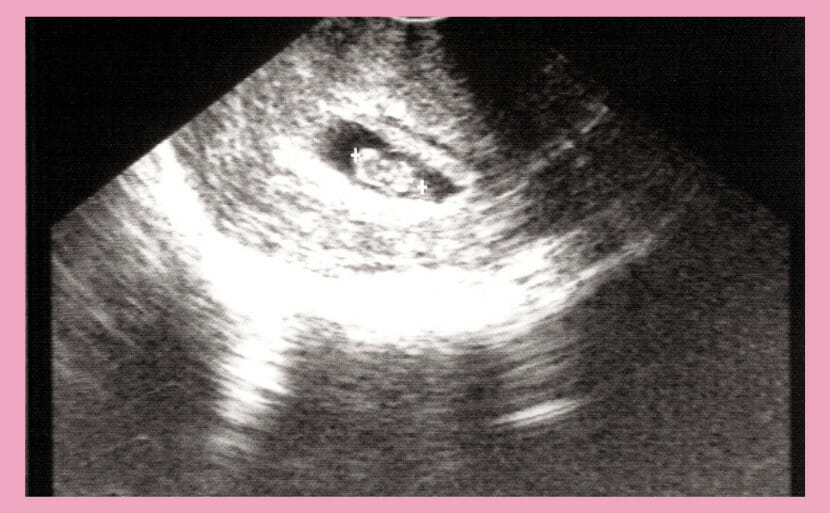
妊娠7週目のエコー写真です。
Characteristics of Down Syndrome seen on ultrasound during the second trimester of pregnancy
During an ultrasound (echo image) taken around the middle of pregnancy (18-22 weeks), it becomes possible to observe the baby’s various organs. In addition to the large head and short limbs seen in the early stages, other characteristics such as congenital heart disease and cleft lip and palate can be seen.
If Down syndrome is suspected based on an ultrasound image, NIPT (non-invasive prenatal testing) is recommended.
It is possible to observe the characteristics of Down Syndrome through an ultrasound examination .
However, the results do not conclusively diagnose Down syndrome .
Pregnant women and their partners will likely consider whether to undergo a maternal serum marker test (quad test), which involves taking a sample of the pregnant woman’s blood, NIPT (non-invasive prenatal testing) , or an amniocentesis test to make a more definitive diagnosis.
If Down syndrome is suspected based on ultrasound images taken early in pregnancy , we recommend undergoing NIPT (non-invasive prenatal testing) to confirm whether there is indeed a possibility of chromosomal abnormalities, including Down Syndrome.
What is NIPT (New Prenatal Testing)?
NIPT (non-invasive prenatal testing) is a test method that analyzes fragments of the baby’s DNA contained in the mother’s blood to determine whether or not there are chromosomal abnormalities. It is characterized by its high accuracy, with a detection rate of 99% (negative predictive value of 99.99%). A high negative predictive value means that if you are tested and told that there is no specific abnormality (such as trisomy 21), the probability that the baby actually has that abnormality is extremely low.
However, NIPT (New Prenatal Testing) is only a screening test to determine whether there is a possibility of disease. For a definitive diagnosis, an amniocentesis must be performed.
Some facilities may have age and other conditions for undergoing NIPT (non-invasive prenatal testing) , but Hiro Clinic NIPT does not impose age restrictions on the test, based on the American College of Obstetrics and Gynecology’s guidelines for NIPT (non-invasive prenatal testing) .
Testing method for NIPT (new type prenatal testing)
NIPT (new type prenatal testing) can be done by taking blood from the mother’s arm. It is a safe test with no risk of miscarriage.
NIPT (new type prenatal testing) test costs
The drawback of NIPT (new type prenatal testing) is that it is not covered by health insurance and must be paid for out of pocket, making it expensive. The cost of the test varies depending on the medical institution and the type of test, but is roughly between 50,000 and 240,000 yen.
At Hiro Clinic NIPT , we offer a variety of courses to suit your needs and budget, so please feel free to consult us when making a reservation.
Please note that prenatal diagnosis is not eligible for medical expense deductions. This is because medical expense deductions are limited to “medical procedures involving diagnosis and treatment,” and prenatal diagnosis, which is merely a test, is not recognized as a medical procedure, just like medical checkups and health checkups.
Things to be aware of when undergoing NIPT (new prenatal testing)
When undergoing NIPT (new prenatal testing) , it is important to pay attention to the deadline for the test. If you are able to undergo a definitive test in the event of a positive result, amniocentesis is required at 15-18 weeks. Therefore , NIPT (new prenatal testing) must produce results before that time, and Hiro Clinic NIPT recommends testing up to the 15th week. By the way , Hiro Clinic NIPT can be performed immediately after pregnancy is confirmed by ultrasound. Reservations can be made as soon as the due date is known.
Probability of Down Syndrome by Age
The probability of having a baby with Down’s syndrome is approximately 1 in 1,000 (0.1%). The exact probability varies depending on the mother’s age at the time of birth, but generally the probability increases with age, and is over 1% for women giving birth at age 40 or older.
Probability of Down Syndrome among women in their 20s
The probability of a pregnant woman in her 20s giving birth to a baby with Down syndrome is extremely low, approximately 1 in 1,667 (0.060%).
Probability of Down Syndrome among women in their 30s
The probability of a woman in her 30s giving birth to a baby with Down syndrome is approximately 1 in 952 (0.105%). For women aged 35 and above, the probability increases to approximately 1 in 378 (0.260%).
Probability of Down Syndrome among women in their 40s
A 40-year-old woman has about a 1 in 106 (0.943%) chance of giving birth to a baby with Down’s syndrome . The chances increase with age, from about 1 in 86 (1.163%) at 41, to about 1 in 50 (2%) at 43, and about 1 in 30 (3.333%) at 45.
After the age of 45, the probability increases dramatically: by age 47, the probability is approximately 1 in 18 (5.556%), and by age 49, the probability is approximately 1 in 11 (9.091% ) .

If your baby is diagnosed with Down Syndrome
If your baby is diagnosed with Down’s syndrome , both parents should first receive a thorough explanation from their doctor. If you decide to give birth, it is a good idea to have a proper evaluation to see if there are any complications that need to be dealt with immediately after birth, and to get in touch with local parent groups (such as the Japan Down Syndrome Association) that provide support for children with Down’s syndrome and their families.
Unfortunately, if you choose to have an abortion, the procedure must be performed before 22 weeks of pregnancy.
summary
Above, we have reviewed the characteristics of Down syndrome , as well as whether Down syndrome can be diagnosed based on an ultrasound (ultrasound photo) taken in the 7th week of pregnancy .
Even if the characteristics of babies with Down syndrome described in this article are seen on an ultrasound, it does not mean that the baby has Down syndrome based on those characteristics alone.
Once the doctor who performed the test has told you about the characteristics of Down syndrome , be sure to receive a thorough explanation of what the test results mean and what subsequent tests you can take if you wish.
The next tests include maternal serum marker test (quad test), NIPT (non-invasive prenatal testing) , and amniocentesis.
Please discuss the matter thoroughly with your partner, and if you find it difficult to decide whether or not to get tested, please consult your doctor and you can receive professional genetic counseling.
When you become pregnant, it’s natural to want to give birth to a happy and healthy baby.
Because of these feelings, you may be upset if an ultrasound scan at a checkup reveals characteristics of Down syndrome in your baby, but there is no need to worry unduly.
If characteristics of Down syndrome are found, the best next step is to consult a doctor or genetic counselor who can properly evaluate the test results and provide detailed information about future testing.
The most important thing is whether the pregnant woman and her partner can decide with confidence whether or not to take the test, after consulting with a specialist.
If you are worried about Down syndrome , especially if you are giving birth at an older age , make good use of ultrasound testing and NIPT (non-invasive prenatal testing) to ensure you are prepared to give birth without anxiety.
【References】
- Today’s Clinical Support – Down Syndrome (Pediatrics)
Article Editorial Supervisor

Dr Hiroshi Oka
NIPT specialist clinic, MD
Graduated from Keio University, School of Medicine
 中文
中文


