13 trisomy, also known as Patou syndrome, is a congenital chromosomal abnormality that can occur in 1 in 5,000 to 12,000 children.
What is Trisomy 13 (Patau Syndrome)?
Patau syndrome, also called trisomy 13 syndrome, is also commonly referred to as trisomy 13. The ‘Patau’ in Patau syndrome is taken from the name of a person, after whom these syndromes were identified as genetic diseases.
In Patau syndrome, one of the autosomes (chromosomes other than the sex chromosome that determines gender) in the cell has one more chromosome (trisomy) than it should have two (chromosome 13), or a part of the other chromosome is duplicated. It is said to be a congenital chromosome disorder caused by a change in the number or an imbalance in the amount of these chromosomes.
Human cells contain 46 chromosomes, which are arranged in sets of two chromosomes each. We receive one set of autosomes (chromosomes 1 through 22) from each of our mother and father. The male sex chromosome is ‘XY’ and the female sex chromosome is ‘XX’.
Other trisomy syndromes similar to Patau syndrome are Trisomy 21 (Down syndrome) and Trisomy 18 (Edwards syndrome).
These trisomies differ not only in facial features and symptoms, but also in life expectancy. Trisomy 13 (Patau syndrome) has the shortest life expectancy among these three trisomies.
Characteristics of Trisomy 13 (Patau syndrome)
The growth and developmental characteristics of Patau syndrome include a small stature and very slow growth and development since fetal period.
In terms of physical characteristics, the child may be born with external features on the face and brain, such as cleft lip or palate (the roof inside the mouth), or have heart disease. It is estimated that about 80% of babies with Patau syndrome have serious heart disease. In addition, a high frequency of babies with Patau syndrome have distinctive symptoms of the reproductive organs, such as testes that are not in their normal position in baby boys and abnormalities in the shape of the uterus, such as a bicornuate uterus in baby girls.
In early childhood, apneic attacks, prolonged periods of no breathing, are common.
Patau syndrome is also associated with relatively severe intellectual disability.
Probability of Trisomy 13 (Patau syndrome)
Approximately 1 in every 5,000 to 12,000 babies born is reported to have Patau Syndrome. About 80% of all cases of Patau syndrome have a complete trisomy 13 ( three autosomal chromosomes 13).
The one extra chromosome, the 13th, is usually inherited from the mother, and the older the mother is, say over 35, the more likely it is that a baby with Patau syndrome might be found.
The probability of detecting a baby with Patau syndrome is 1 in 1,000 in amniotic fluid testing at 16 weeks’ gestation and 1 in 12,000 at birth.
What is of concern is that once a mother conceives a baby with Patau syndrome, there is a possibility that in her next pregnancy, she may conceive a baby with the same Patau syndrome or other chromosomal abnormalities.
Concerning this, there are also reports that a mother who has given birth to a baby with Patau syndrome in the past is unlikely to conceive a baby with a chromosomal abnormality such as Patau syndrome in her next pregnancy.

Causes of Trisomy 13 (Patau Syndrome)
Patau syndrome is caused by the duplication of the entire length or part of the chromosome 13, as described above, resulting in an increase in the number of chromosomes in the cell and an imbalance in the amount of genes, causing various genes to fail to function properly, resulting in the characteristic birth defects.
Which begs the question, why would the number of chromosomes in the chromosome 13 be increased?
One reason for this is that the chromosomes were not successfully separated during the fertilization process between sperm and egg.
In the cell before the egg and sperm are fertilized, the chromosomes are supposed to be divided into sets of 23 chromosomes, but for some reason, the two 13th largest chromosomes have entered the egg or sperm together.
Other cases of ‘translocation’ also occur, where two different chromosomes are each shredded in the step before the chromosome is divided into 23 chromosomes, and the two chromosome fragments are exchanged and attached to each other. This can result in ‘partial trisomy,’ in which only a portion of the 13th chromosome is trisomic.
This inability of the chromosomes in the gametes to separate properly during the process of sperm and egg division and fertilization results in trisomy, and the tendency for these to occur is also said to be related to the mother’s age.
Symptoms of Trisomy 13 (Patau Syndrome)
Patau syndrome is often associated with complications such as delayed growth and development from the fetal period, various physical characteristics, and various heart diseases.
Other congenital features and diseases are also present as symptoms of Patau syndrome, including:
Physical Characteristics
- Microcephaly (delayed brain development)
- Scalp defects
- Partial skull defect (partial loss of skull)
- Small eyes, monocular (only one eye), anophthalmia
- Spacing between the two eyes is open
- Low nasal bridge
- Iris coloboma (loss of part of the iris that controls light entering the eye)
- Defects in the pupil (the colored part of the eye)
- Poor development of the retina (the transparent structure at the back of the eye that senses light)
- Cleft lip and palate, high palate
- Deformity of the ears
- Ears in a low position
- Sagging skin on the back of the neck
- Deep wrinkles in a straight line on the palms of the hands
- Extra fingers on the hands and feet
- Hand grip with fingers overlapping other fingers
- Protruding heels
- Poor nail development
Complications Related to the Central Nervous System
- Total forebrain folliculopathy (failure of the left and right cerebral hemispheres = forebrain to separate properly and remain as one)
- Convulsions
Complications Related to the Respiratory System
- Apneic Attacks (prolonged periods of no breathing)
- Pharyngeal and Tracheal Softening
Congenital Heart Diseases
- Ventricular Septal Defect (a hole in the wall between the left and right ventricles)
- Open Ductus Arteriosus (the duct connecting the aorta to the pulmonary artery remains open)
- Atrial Septal Defect (a hole in the wall between the left and right atria)
Complications Related to the Urinary System
- Abnormalities in the shape of the kidneys
- Inguinal hernia (intestines and visceral fat protruding through thin abdominal muscles)
- Small Penis
- Retained Testicle (testicle is not in its normal position)
- Abnormal Shape of the Uterus (e.g., bicornuate uterus that divides into two parts)
Complications Related to the Endocrine System
- Hypothyroidism
- Prolonged Hypoglycemia (severe hypoglycemia)
Hematological Abnormalities
- Abnormal shape of white blood cells called neutrophils, which produce a defensive response to invading agents such as bacteria
Other Complications
- Hearing Impairment
- Intellectual and Developmental Disabilities
- Gastroesophageal Reflux Disease (acid in the stomach flows back into the esophagus)
- Umbilical Cord Hernia (the stomach, intestines, liver, etc. remain protruding inside the umbilical cord)
- Common Bile Duct Dilatation (dilation of the bile ducts, resulting in bile stasis)
- Biliary Stasis
The symptoms of these features and diseases are different for each baby. However, among the most common are severe mental disability, facial and foot characteristics, heart disease and hearing loss, developmental delays, and apneic seizures.
Testing for Trisomy 13 (Patau Syndrome)
Testing to confirm the diagnosis of Patau Syndrome can be performed before or after the baby is born.
Prenatal: Ultrasound, Testing using the Mother’s Blood
Trisomy 13 can also be detected on prenatal ultrasound examination by the following developmental delays and physical characteristics of the baby:
- IUGR (Intrauterine Growth Restriction)
- Total anterior encephalocele
- Cleft lip and palate
- Microcephaly
- Microphthalmia
- Monocular disease
- Heart abnormalities (ventricular septal defect, atrial septal defect, patent ductus arteriosus)
- Renal cysts (spherical pouches seen in the kidneys)
- Hyperamniotic fluid ( excessive amount of amniotic fluid)
- Polydactyly ( excess number of fingers)
- Single umbilical artery (only one umbilical artery)
- Hernia of the umbilical cord (hernia of the umbilical cord)
If these findings are observed on ultrasound, the next step is NIPT (New Non-invasive Prenatal Testing) conducted using the expectant mother’s blood.
If Patau syndrome is suspected, or if the results are unclear, an amniotic fluid testing and chorionic villus test will be performed to determine if there are any other chromosomal abnormalities found in the baby.
Chromosome examination tests include the fluorescence in situ hybridization (FISH) method and chromosome microarray examination (CMA), which can be used to identify chromosomes and examine deletions of smaller chromosome parts.
Therefore, if it is found that there are three chromosomes 13, a definite testing for Patau syndrome will be performed. Before and after a definitive diagnosis is made and before and after undergoing testing, extensive informational and genetic counseling is advised.

After Birth: Assessment of Physical Characteristics and Testing with the Baby’s Blood
After the baby is born, you may initially suspect Patau syndrome based on physical characteristics/appearance, and testing with the baby’s blood will then confirm the suspected diagnosis.
Other tests include cardiac ultrasound to check for congenital abnormalities of the heart, head ultrasound and MRI to check the brain, and X-ray and CT scans to check for abnormalities in the gastrointestinal tract and kidneys.
Treatments for Trisomy 13 (Patau Syndrome)
There is currently no fundamental treatment for the complications that babies with Patau syndrome have. Supportive care, such as easing conditions, is used to treat the complications seen in each baby.
In some cases, surgery is performed to treat a heart condition that is present at birth.
Other supportive treatments include administration of oxygen, ventilators, intravenous fluids, and oral medications, depending on the symptoms observed. If the child is unable to be fed by mouth, tube feeding or gastric banding may be considered.
In choosing any of these treatment options, it is important to discuss the treatment plan with the family, respect their wishes, and provide them with sufficient information about the prognosis and the risks of treatment and surgery.
Prognosis of Trisomy 13 (Patau Syndrome)
Support from the medical, psychological, and welfare aspects to help babies and their families have a better life together is important.
The following database links from the Japanese Society for Gene Therapy can be checked (http://www.gene-dt.jp/frame/f_DB.html) for the websites of the departments in charge of gene therapy, genetic counseling, etc. at major medical institutions.
There are also associations that support families with children with Patau syndrome, such as the ‘Parents’ Association to Support Children with 13 Trisomy’ (http://www.13trisomy.com/next.html).
The actual frequency of babies born with Patau syndrome tends to be low, and in some respects it has been difficult to track cases to determine what kind of care is needed.
Recently, however, advances in pediatric care have led to an increase in the number of cases of children being discharged from hospital NICUs ( Neonatal Intensive Care Units) and being cared for at home, and there seems to be a trend toward medical intervention for children with Patau syndrome.
At home, respiratory support and respiratory care are necessary, such as the use of a ventilator for children with frequent breath-holding seizures or tracheostomies, as well as epilepsy care, and it is also important to have a visiting physician who can examine the patient at home for their condition.
Summary
Patau syndrome is a chromosome abnormality caused by an extra autosomal chromosome 13 instead of the two. The older the mother is, the more likely it is to occur.
Babies grow normally in the uterus, although sometimes with developmental delays, and require early detection through Ultrasound and NIPT (New non-invasive Prenatal Testing) as well as coping and treatment after birth. Even though there is currently no fundamental cure, it is essential to provide sufficient explanation and a supportive medical, welfare, and rehabilitation system for parents in advance when undergoing ultrasound, prenatal diagnosis, amniotic fluid testing, chorionic villus examination, and supportive care after birth, as well as during treatment.
13 trisomy, also known as Patou syndrome, is a congenital chromosomal abnormality that can occur in 1 in 5,000 to 12,000 children.
Article Editorial Supervisor
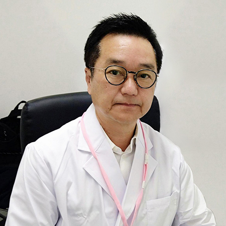
Dr. Shun Mizuta
Head Doctor, Hiro Clinic NIPT Okayama
Board Certified Pediatrician, Japan Pediatric Society
As a pediatrician, he has been engaged in community medicine in Okayama Prefecture for nearly 30 years.
Currently, he is working to educate the community about NIPT as the Head Doctor of Hiro Clinic NIPT Okayama, utilizing his experience as a pediatrician.
Brief History
1988 – Graduated from Kawasaki Medical University
1990 – Clinical Assistant, Kawasaki Medical University, Department of Pediatrics
1992 – Department of Pediatric Neurology, Okayama University Hospital
1993 – Head of the First Department of Pediatrics, Ihara Municipal Hospital, Ihara City
1996 – Mizuta Kodomo Hospital
Qualifications
Board Certified Pediatrician
 中文
中文


