Learn about the process and technology behind Non-Invasive Prenatal Testing (NIPT) and how it's performed. Discover its benefits and implications for expecting parents.
Introduction
NIPT (Non-Invasive Prenatal Testing) is a test that examines chromosomal abnormalities such as Trisomy 21 (Down syndrome), Trisomy 18 (Edwards syndrome), and Trisomy 13 (Patau syndrome) in the fetus from blood collected from the mother. Compared to conventional prenatal diagnosis using blood, the accuracy of the test itself is considered very high in terms of sensitivity and specificity. The blood of pregnant women contains DNA fragments derived from the baby. NIPT examines the possibility of these chromosomal abnormalities in the baby by analyzing DNA fragments derived from the baby.
As for the advantages of NIPT, unlike the amniocentesis and chorionic villus sampling, which have been performed as prenatal diagnoses so far and require needle insertion into the uterus, NIPT is performed by taking blood from the mother’s arm, making it less invasive (less damage to the body) for the mother and eliminating the risk of miscarriage (0.3-1%) associated with these tests. Furthermore, due to the high accuracy of NIPT, the obtained test results can be used by doctors as a reliable indicator to assess the possibility of disease risk.
However, as a caution regarding NIPT, it is important to understand that NIPT is a type of screening test and not a definitive test that provides a conclusive answer. It only examines the probability of whether the fetus has a high or low risk of chromosomal abnormalities. Therefore, before undergoing NIPT testing, it is recommended to consult with a physician thoroughly regarding the chromosomal abnormalities that can be detected by NIPT, the principles of NIPT, the mechanism of the test, as well as its advantages and cautions.
In this article, we will introduce from the principle of NIPT to how the blood collected from the mother is tested and analyzed to obtain the results of NIPT testing, as well as the considerations for NIPT.
Principle of NIPT (Non-Invasive Prenatal Testing)
The principle of NIPT (Non-Invasive Prenatal Testing) is to collect fragments of fetal DNA present in the mother’s blood during pregnancy and analyze those DNA fragments in detail using a machine (sequencer) to test for the possibility of the fetus having chromosomal disorders such as trisomy 21, trisomy 18, trisomy 13, etc. To confirm whether the fetus, not the pregnant woman, has chromosomal abnormalities and to test the possibility of the fetus being affected by these chromosomal disorders, it is necessary to analyze DNA derived from the fetus rather than from the mother, so fetal DNA in maternal blood becomes the target of NIPT testing. In this section, we will introduce the DNA, sequencer, and analysis methods necessary for NIPT testing.
DNA Required for NIPT (Non-Invasive Prenatal Testing)
In conventional prenatal diagnosis, amniotic fluid or chorionic villus samples containing fetal DNA are collected, and the sequence of fetal DNA is examined to determine whether chromosomal disorders are present.
On the other hand, NIPT (Non-Invasive Prenatal Testing) utilizes fetal DNA present in the mother’s blood. Normally, DNA is stored as chromosomes within the nucleus of a single cell in the body. However, it is known that DNA fragments also flow into the bloodstream, and these are referred to as cell-free DNA (cfDNA). In NIPT testing, cfDNA is utilized. Not only in NIPT but also in the testing of cancer and other diseases, cfDNA is used. The process from cfDNA collection to diagnosis is considered one of the highly reliable and safe testing technologies.
The cfDNA used in NIPT testing originates from the fetal blood of the placenta flowing into the mother’s bloodstream. Testing facilities extract cfDNA and analyze its DNA sequence. Based on the results obtained, doctors determine the possibility of the fetus having chromosomal disorders before birth.

Sequencer
A sequencer refers to a machine that reads the sequence of DNA, and currently, there are the latest ones called Next Generation Sequencers (NGS). Sequencers, which originally used a method called the Sanger method in the 1970s, have the same basic mechanism even with NGS, which aims to read the sequence of extracted DNA bases.
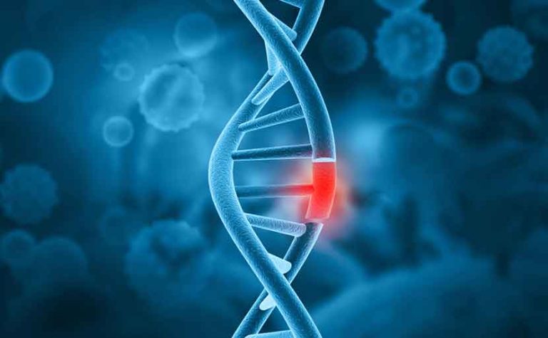
DNA of living organisms is formed by sequences of four types of bases: A (adenine), T (thymine), G (guanine), and C (cytosine). Additionally, bases are fundamentally paired, with A pairing with T and G pairing with C (complementarity), forming a state where two DNA strands are bound together (double-stranded DNA). These strands fold to form structures called chromosomes, which are stored within the nucleus of each cell. Specific sites within the DNA have common base sequences, and reading whether they are normal sequences (sequencing) identifies genetic abnormalities.
The specific method of a sequencer involves initially extracting and purifying the desired DNA from a sample, putting it into a single-stranded state. Then, reagents called primers, which have specific base sequences, bind to the ends of the target DNA sequence. Subsequently, a mixture of reagents called ddNTPs, each tagged with a different fluorescent color corresponding to A, T, G, or C, along with a reagent called DNA polymerase, work together to sequentially add bases to the DNA strand, reading the original extracted DNA’s gene sequence.
However, traditional sequencing using conventional sequencers faced two main challenges. Firstly, it could only read one DNA strand per sequencing run. To find multiple genetic abnormalities, multiple DNA extractions and sequencings were necessary, consuming much time and effort. Secondly, traditional sequencing required a large amount of DNA, meaning substantial samples were needed at the extraction stage. This posed difficulties, especially for reading low amounts of DNA, such as cfDNA, requiring significant amounts of blood or tissue, which was highly invasive and challenging.
To address these challenges, Next Generation Sequencing (NGS) was developed. For the first challenge, NGS creates a catalog of DNA, known as a DNA library, within the inspection process, allowing for the simultaneous reading of DNA base sequences at levels ranging from millions to billions in a single sequencing run. Essentially, NGS enables sequencing at a level comparable to performing traditional sequencing over a million times in one go. Regarding the second challenge, the development of reagents and methods for more efficient and abundant DNA extraction has contributed. With NGS, even small amounts of DNA can be sequenced efficiently, making it possible to detect genetic abnormalities even with low amounts of DNA, such as cfDNA.
Analysis Methods
There are various methods for analyzing fetal-derived cell-free DNA (cfDNA). Apart from the commonly used SNP analysis (single nucleotide polymorphism analysis, which examines whether one base is different, for example, if a position that should have been A is T), and microarray analysis (where multiple DNA sequences are arranged on a chip and the DNA from the sample being examined is bound to them to detect DNA amounts), the massively parallel sequencing (MPS) method is considered standard.
The MPS method combines NGS analysis using fetal-derived cfDNA with the analysis of component amounts derived from each chromosome. In other words, it quantitatively examines which chromosomes the obtained fetal-derived cfDNA originated from. For example, in a case where there’s a high likelihood of trisomy 21 in the fetus, the result may show three copies of chromosome 21 instead of the normal two, indicating that the amount of DNA derived from chromosome 21 is 1.5 times higher (3÷2=1.5).
However, whether in NIPT (non-invasive prenatal testing) or other genetic tests to confirm genetic disorders, it’s important to recognize that none of these analysis methods provide 100% confirmation. They should be considered as screening tests rather than definitive diagnostic tools.
Mechanism of NIPT (Non-Invasive Prenatal Testing)
NIPT (Newborn Screening Test) works roughly as follows:
Blood collection from pregnant women → Extraction of fetal-derived cell-free DNA (cfDNA) → Library preparation → Sequence analysis of cfDNA → Determination of the possibility of chromosomal abnormalities.
Maternal Blood Collection
Specifically, first, about 10 to 20 mL (roughly 2 to 4 teaspoons) of blood is drawn from the arm of a pregnant woman whose pregnancy has been confirmed by ultrasound examination. Fetal-derived cell-free DNA (cfDNA) floating in the blood is then extracted. At this stage, it’s important to note that maternal-derived cfDNA is also included, which will be addressed later.
Why Blood Collection is Necessary
The reason for blood collection from the mother is that it’s a less invasive method compared to other techniques such as collecting amniotic fluid or chorionic villus sampling.
While obtaining tissue or cells directly from the fetus would theoretically provide the highest testing accuracy when collecting fetal-derived DNA, direct sampling poses significant risks to the fetus and may lead to a risk of miscarriage for both the mother and the fetus. Therefore, by extracting fetal-derived cell-free DNA (cfDNA) from the mother’s blood, prenatal diagnosis becomes possible without impacting the fetus, thus avoiding potential risks associated with direct sampling.
Maternal Blood Centrifugation
To extract fetal-derived cfDNA from maternal blood, the maternal blood is separated into its components using a device called a centrifuge.
Human blood is naturally divided into two main components: the liquid component (plasma, about 55%) and cellular or protein components and other substances, all of which flow throughout the body in a mixed state. By centrifuging the blood collected in a blood collection tube, it is separated into layers of plasma, buffy coat (a thin layer containing white blood cells), and red blood cells. To give an analogy, it’s like putting water and oil in the same container and letting them settle; since water is heavier than oil, the oil sinks to the bottom. This separation technique is commonly used in hospitals and health check-ups for blood tests.
Among the blood components, fetal-derived cfDNA is contained within the plasma, while the buffy coat and red blood cells are of maternal origin. Therefore, to conduct accurate NIPT (non-invasive prenatal testing), during the preparation stage of the test, it is crucial to carefully separate and isolate only the plasma.
Analysis of Results
The analysis of the results begins by first collecting all DNA fragments present in the maternal plasma and preparing a library with special labels attached. This library is then sequenced using NGS (Next Generation Sequencing), specifically the MPS (Massively Parallel Sequencing) method. During the analysis steps, each DNA fragment is identified to determine its origin on which chromosome.
The genetic information obtained from the DNA fragments is aligned on a reference sequence, and the abundance of genetic information at the relevant loci is compared to cutoff values (criteria for determining normal and abnormal for chromosomes 21, 18, and 13). This comparison helps in determining whether the result is positive, negative, or requires further evaluation.
Hiro Clinic NIPT tests vary depending on the plan, but for 95% of cases, results are available within 8 days.
These steps are explained step by step.
Sequencing
As mentioned earlier, in NIPT (non-invasive prenatal testing), NGS (Next Generation Sequencing) is used to sequence fetal-derived cfDNA. Since maternal plasma also contains maternal-derived cfDNA (some estimates suggest that around 10% of maternal plasma cfDNA is of fetal origin at around 10 weeks of pregnancy), both maternal and fetal-derived cfDNA are sequenced comprehensively together.
Specifically, the process involves watching an explanatory video about NIPT at the clinic, filling out a questionnaire and consent form, undergoing a consultation with a doctor, and then blood collection. Subsequently, either at the clinic or at an accredited facility, maternal plasma is obtained through centrifugation, and cfDNA is extracted from maternal plasma for comprehensive sequencing using NGS.
Identification
After the comprehensive sequencing (MPS method) is conducted, the process moves to the identification step. As mentioned in the analysis method section, by quantitatively measuring which chromosome the fetal-derived cfDNA originates from, it becomes possible to detect abnormalities such as trisomies of chromosomes 21, 18, and 13, as well as abnormalities in the sex chromosomes (biologically, males have one X and one Y chromosome, while females have two X chromosomes). Additionally, gender determination can also be performed.
Evaluation
NIPT results are determined quantitatively, as mentioned in the identification section. Since DNA is not visible to the naked eye, data must be quantified and clear criteria established.
Specifically, the data obtained from NGS is compared to cutoff values for determination. Cutoff values can be understood simply as reference values. By establishing specific numerical values as cutoff values, determinations are made based on how far the obtained values deviate from these reference values.
For instance, in a health check-up, when results are returned, there are often “reference values” provided as a range. If the results fall outside of this range, it may be labeled as “requires further testing,” and so on. Similarly, in NIPT, cutoff values are established, and determinations are made by comparing the obtained values against these reference values.

Considerations for NIPT (Non-Invasive Prenatal Testing)
NIPT (Non-Invasive Prenatal Testing) is one of the non-definitive tests (which also includes maternal serum marker tests) that determine the likelihood of having a chromosomal abnormality in a fetus probabilistically rather than conclusively before birth. Therefore, it is important to undergo confirmatory diagnosis if your fetus is deemed likely to have a chromosomal abnormality based on NIPT results. To undergo confirmatory diagnosis, it is necessary to undergo definitive tests (amniocentesis, chorionic villus sampling). In addition, there are other important points to consider with NIPT, such as the following:
Chorionic Placental Mosaicism (CPM)
It’s important to note that the fetal-derived cfDNA present in maternal plasma used in NIPT is primarily of placental origin, meaning it doesn’t exclusively represent cfDNA from the fetus. Due to this characteristic, there is a risk that the test results may not accurately reflect the chromosomal information of the fetus in cases where there are differences in chromosomal karyotype between the fetus and the placenta (referred to as chorionic placental mosaicism, or CPM).
Vanishing Twin
In the initial stages of pregnancy, if a woman conceives twins but one fetus dies early in the pregnancy and is absorbed by the uterus, it’s referred to as a vanishing twin phenomenon. This term describes the situation where one fetus vanishes and is not visible on ultrasound or other imaging tests. In such cases, there is a possibility that the DNA of the deceased fetus may still be detected, potentially affecting the results of NIPT.
Cases of “False Positive,” “False Negative,” or “Re-testing (Pending Evaluation)”
Basically, the test results are reported as either “positive” or “negative,” but it cannot be completely ruled out that they may also be categorized as “false positive,” “false negative,” or “retesting (pending determination).”
“False positive” refers to the situation where a fetus is actually healthy but is classified as positive. “False negative” occurs when a fetus actually has a chromosomal abnormality but is classified as negative.
There are various reasons for these results, but they can generally be attributed to biological factors in the mother or fetus, or to conditions such as confined placental mosaicism (CPM) as mentioned earlier. Examples of biological factors in the mother or fetus include the influence of vanishing twin, chromosomal abnormalities that are asymptomatic in the mother, and effects based on the clinical situation of the mother’s symptoms (such as the presence of tumors, medication effects, autoimmune diseases, organ or bone marrow transplants, etc.). Other factors include errors due to the limitations of the test.
Currently, based on domestic research results, it is estimated that the proportion of these results is around 0.1% to 0.3%.
Conclusion
NIPT (Non-Invasive Prenatal Testing) is one of the prenatal diagnostic tests that can be performed from early pregnancy. It is a simple, minimally invasive, and highly accurate test. Furthermore, because it employs cutting-edge technology, further improvements are expected, such as increased accuracy and the possibility of testing with even smaller sample sizes in the future.
For parents-to-be, NIPT (Non-Invasive Prenatal Testing) is considered a valuable test to alleviate concerns about whether their future child has chromosomal abnormalities. However, it’s important to understand that NIPT is ultimately a non-confirmatory test (a screening test), and depending on the results, it may be necessary to undergo invasive confirmatory testing. There are several important considerations to take into account. Therefore, it’s crucial to decide whether to undergo the test after thorough consultation with medical staff such as doctors and counselors.
【References】
- Ministry of Health, Labour and Welfare – NIPT: Noninvasive Prenatal Testing 無侵襲的出生前遺伝学的検査
- ACOG – Non-Invasive Prenatal Testing
- ACOG – Current ACOG Guidance
- ACOG – Prenatal Genetic Screening Tests
- Pre-Natal Examination Accreditation System Operation Committee – To Medical Institutions and Testing Facilities
- Japan Medical Association – Guidelines for Information Provision on Prenatal Testing such as NIPT and Certification of Facilities (Medical Institutions and Testing Facilities)
- Japan Medical Association – Brochure on Non-Invasive Prenatal Genetic Testing
- Japan Medical Association – Non-Invasive Prenatal Genetic Testing
- J-STAGE – Fetal Physiology: Journal of the Japanese Society of Clinical Anesthesiologists, Vol.38 No.4, pp. 538-541, 2018
- Hyogo University of Medicine Hospital Obstetrics and Gynecology Department – Information on Prenatal Diagnosis
- ACOG – From the Basics of NIPT to Future Perspectives
- Illumina – Differences Between NGS and Sanger Sequencing
- Illumina –Targeted next-generation sequencing versus qPCR and Sanger sequencing
- Thermo Fisher Scientific – What is a Microarray? | Principles and Analysis Methods of Gene Expression Analysis Using Microarrays
Q&A
-
QWhat type of prenatal diagnosis is recommended by the Ministry of Health, Labor and Welfare?The Ministry of Health, Labor and Welfare (MHLW) recognizes nonconfirmatory tests such as ultrasonography, maternal serum marker test (Quattro test), and NIPT (new-type prenatal diagnosis), as well as confirmatory tests such as chorionic villus examination and amniotic fluid test. If the non-confirmatory test is positive, a confirmatory test is recommended.
-
QShould I always get a prenatal checkup?Prenatal diagnosis is not necessarily required for all pregnant women.It is important to consider it in consultation with a physician, depending on the circumstances and desires of the individual pregnant woman.It is especially worth considering in cases of older pregnancies and family history.
-
QWhat are the principles of NIPT (new prenatal diagnosis)?NIPT is a method of analyzing fetal-derived DNA fragments (cfDNA) present in the mother's blood to test for the possibility that the fetus has chromosomal abnormalities such as 21-trisomy, 18-trisomy, or 13-trisomy.It is widely used as a highly accurate and non-invasive test.
 中文
中文


